An easy technique for silicophytolith visualization in plants through tissue clearing and immersion oil mounting
DOI:
https://doi.org/10.31055/1851.2372.v54.n3.25359Keywords:
anatomy, dicotyledons, Equisetum sp, immersion oil, monocotyledons, silicophytoliths, tissue clearingAbstract
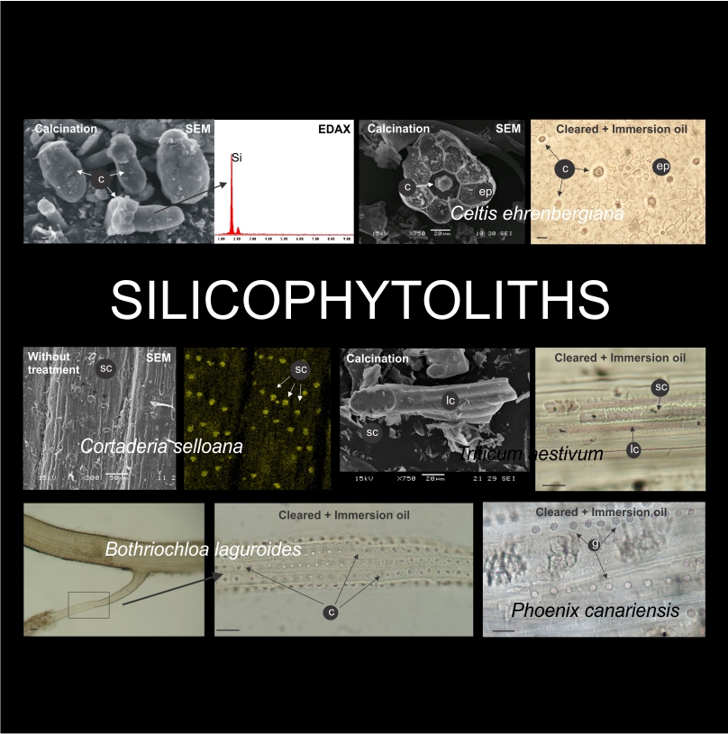
Background and aims: Different methodologies were proposed for the detection of silica deposits (silicophytoliths) in plant tissues. These methodologies include dry and wet ashing (which destroy the surrounding tissue), phenol staining (which is toxic), safranin–crystal violet lactone and fast green–methyl red staining (not specific for silica), fluorescence microscopy, electronic microscopy, EDAX and Raman analyses (which involve expensive equipment). Here we presented an easy and cheap method based on tissue clearing and immersion oil mounting.
M&M: We tested the methodology in longitudinal and cross sections of culms, leaves and roots of ten species that effectively accumulate silica. We applied different clearing techniques according to the type of plant material, we mounted in immersion oil and observed under light microscope. We compared the results with the ones obtained by traditional silicophytolith techniques (dry ashing, phenol staining and SEM-EDAX analyses).
Results: Silica deposits were observed in all species and organs analyzed, and the observations were coincident with the results obtained by other techniques. It was also possible to identify calcium crystals, allowing the description of the most common biomineralizations produced by plants.
Conclusions: The technique here proposed can be used for exploratory as well as for specific studies about the content and distribution of silicophytoliths in almost any tissue, organ and plant species. It can be applied in any laboratory, because it does not require expensive or hardly available equipment.
Downloads
Downloads
Published
How to Cite
Issue
Section
License
Provides immediate and free OPEN ACCESS to its content under the principle of making research freely available to the public, which fosters a greater exchange of global knowledge, allowing authors to maintain their copyright without restrictions.
Material published in Bol. Soc. Argent. Bot. is distributed under a Creative Commons Attribution-NonCommercial-ShareAlike 4.0 International license.