
La anatomía de la raíz como evidencia filogenética para los helechos Cheilanthoides (Pteridaceae)
DOI:
https://doi.org/10.31055/1851.2372.v59.n3.44811Palavras-chave:
Anatomía radicular, corteza radicular, endodermis, filogenia, helechos cheilanthoidesResumo
Introducción y Objetivos: A nuestra hipótesis sobre las relaciones evolutivas de los helechos cheilanthoides, fundamentada en datos moleculares, y parcialmente por datos morfológicos y reproductivos, aquí se suma un estudio de la anatomía de la raíz en los géneros de la subfamilia con presencia en América del Sur.
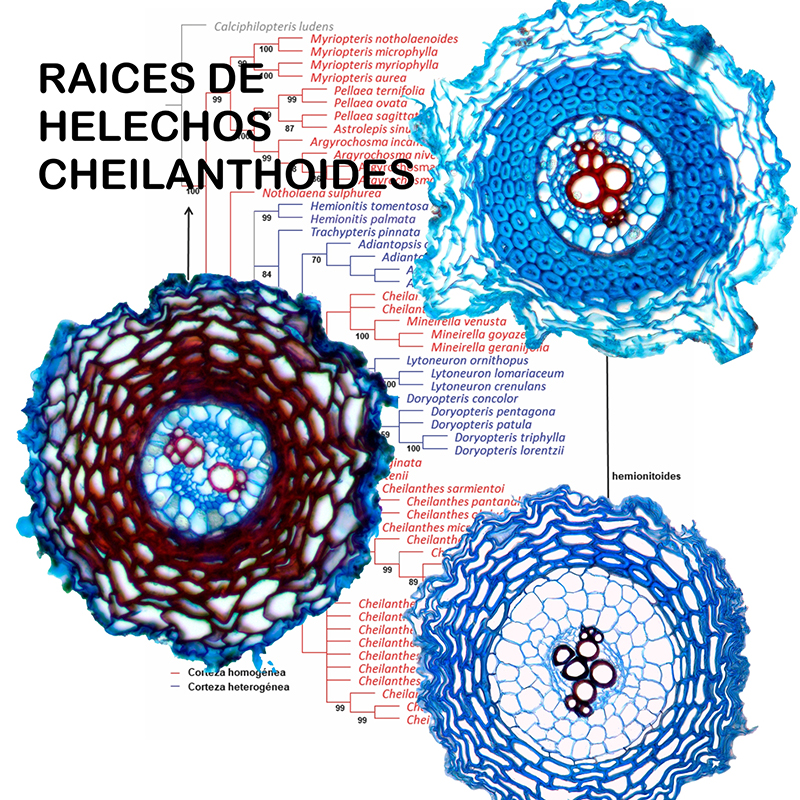
M&M: Se analizaron anatómicamente caracteres radiculares en 48 especies sudamericanas representando los 12 géneros de Cheilanthoideae con presencia en América del Sur. Para el análisis anatómico y caracterización de los tejidos se aplicaron técnicas de histología vegetal. Se identificaron caracteres con posible valor diagnóstico y se mapearon sobre una filogenia molecular de Cheilanthoideae basada en tres marcadores plastidiales (rbcL, trnL-F y rps4).
Resultados: El carácter “tipo de corteza radicular” mostró una distribución de estados que se ajustó a los principales clados o géneros de la filogenia de la subfamilia. El estado corteza homogénea resultó plesiomórfico para Cheilanthoideae, mientras que el estado corteza heterogénea habría sido adquirido por algunos géneros del clado hemionitoide: Hemionitis y los géneros del clado Adiantopsis-Doryopteris (excepto Mineirella). Adicionalmente, discutimos el rol de los diferentes tejidos en la mecánica de las raíces de este grupo.
Conclusiones: El carácter “tipo de corteza” podría contribuir a la caracterización de géneros y la asignación genérica de especies confusas en la clasificación de Cheilanthoideae. Sin embargo es necesaria la resolución completa de las relaciones filogenéticas dentro del clado hemionitoide en la filogenia global de la subfamilia.
Referências
ANDERSEN, T. G., D. MOLINA, J. KILIAN, R. B. FRANKE, … & N. GELDNER. 2021. Tissue-Autonomous Phenylpropanoid Production Is Essential for Establishment of Root Barriers. Curr Biol 31: 965-977. https:--doi.org-10.1016-j.cub.2020.11.070
BRUNO, G., L. STIEFKENS, M. HADID, I. LISCOVSKY, … & N. DOTTORI. 2007. Efecto de la contaminación ambiental en la anatomía de la hoja de Ligustrum lucidum (Oleaceae). Bol. Soc. Argent. Bot. 42: 231-236. https:--doi.org-10.1086-337646
CHAPPLE, C. C. S. & R. L. PETERSON. 1987. Root Structure in the Fern Platycerium bifurcatum (Cav.) C. Chr. (Polypodiaceae). Bot. Gaz. 148: 180-187.
D’ AMBROGIO DE ARGÜESO, A. 1986. Manual de técnicas en histología vegetal. Hemisferio Sur S.A., Buenos Aires.
DAMUS, M., R. L. PETERSON, D. E. ENSTONE & C. A. PETERSON. 1997. Modification of cortical cell walls in roots of seedless vascular plants. Bot. Acta 110: 190-195.
https:--doi.org-10.1111-j.1438-8677.1997.tb00628.x
DUBROVSKY, J. G. & S. SHISHKOVA. 2013. Developmental adaptations in roots of desert plants with special emphasis on Cacti. En: ESHEL, A. & T. BEECKMAN (eds.), Plant Roots the Hidden Half. 4th Ed., vol. 28: 1-13. CRC Press-Taylor & Francis Group, Boca Raton.
ENSTONE, D. E. & C. A. PETERSON. 2005. Suberin lamella development in maize seedling roots grown in aerated and stagnant conditions. Plant Cell Environ. 28: 444-455.
https:--doi.org-10.1111-j.1365-3040.2005.01286.x
ENSTONE, D. E. & C. A. PETERSON & F. MA. 2003. Root endodermis and exodermis: structure, function, and responses to the environment. J. Plant Growth Regul. 21: 335-351.
https:--doi.org-10.1007-s00344-003-0002-2
FELSENSTEIN, J. 1985. Phylogenies and the comparative method. Amer. Naturalist 125: 1-15.
https:--doi.org-10.1086-284325
GELDNER, N. 2013. The Endodermis. Annu. Rev. Plant Biol. 64: 531-558.
https:--doi.org-10.1146-annurev-arplant-050312-120050
GOLOBOFF, P., J. FARRIS & K. C. NIXON. 2008. TNT, a free program for phylogenetic analysis. Cladistics 24: 774-786. https:--doi.org-10.1111-j.1096-0031.2008.00217.x
HERNÁNDEZ, M. A. & F. P. RODRÍGUEZ. 2010. Morfología y anatomía del esporofito de Adiantopsis
chlorophylla (Pteridaceae). Lilloa 47: 85-94.
HERNÁNDEZ, M. A., G. TERÁN. & P. L. ALBORNOZ. 2010. Morfología, anatomía y endomicorrizas en el esporofito de Doryopteris concolor (Pteridaceae). Lilloa 47: 74-84.
HERNÁNDEZ, M. A, M. G. NÁZARO & M. E. ORQUEDA. 2011a. Morfología y anatomía del esporofito de Cheilanthes pilosa (Pteridaceae). Lilloa 48: 74-82.
HERNÁNDEZ, M. A., O. VARELA, Y. E. FERNÁNDEZ & M. G. NADRA. 2011b. Caracterización morfológica y anatómica del esporofito de Trachypteris pinnata (Pteridaceae) en relación con la xeromorfía. Lilloa 48: 153-165.
HERNÁNDEZ, M. A., L. TERÁN, M. MATA, O. G. MARTÍNEZ & J. PRADO. 2014. Helical Cell Wall Thickenings in Root Cortical Cells of Polypodiaceae Species from Northwestern Argentina. Amer. Fern J. 103: 225-240. https:--doi.org-10.1640-0002-8444-103.4.225
HOSE, E., D. T. CLARKSON, E. STEUDLE, L. SCHREIBER & W. HARTUNG. 2001. The exodermis: a variable apoplastic barrier. J. Exp. Bot. 52: 2245-2264. https:--doi.org-10.1093-jexbot-52.365.2245
HOU G., J. P. HILL, E. B. BLANCAFLOR. 2004. Developmental anatomy and auxin response of lateral root formation in Ceratopteris richardii. J. Exp. Bot. 55: 685-693.
KENRICK, P. & P. R. CRANE. 1997. The origin and early evolution of plants on land. Nature 389: 33-39. https:--doi.org-10.1038-37918
KETELAAR, T. & A. M. C. EMONS. 2009. The actin Cytoskeleton in Root Hairs: a cell elongation device. En: EMONS, A. M. C. & T. KETELAAR (eds.), Root hairs. Plant Cell Monographs Ed., vol. 12: 211-232. Springer, Berlin, Heidelberg. https:--doi.org-10.1007-978-3-540-79405-9_8
LEROUX, O., A. BAGNIEWSKA-ZADWORNA, S. K. RAMBE, J. P. KNOX, … & R. L. L. VIANE. 2011. Non-lignified helical cell wall thickenings in root cortical cells of Aspleniaceae (Polypodiales): histology and taxonomical significance. Ann. Bot. 107: 195-207. https:--doi.org-10.1093-aob-mcq225
LI, F. W., K. M. PRYER & M. D. WINDHAM. 2012. Gaga, a new fern Genus segregated from Cheilanthes (Pteridaceae). Syst. Bot. 7: 845-860. https:--doi.org-10.1111-jse.12723
LINK-PÉREZ, M. A., L. E. WATSON & R. J. HICKEY. 2011. Redefinition of Adiantopsis Fée (Pteridaceae): Systematics, diversification, and biogeography. Taxon 60: 1255-1268.
LÍŠKA, D., MARTINKA, M., KOHANAVA, J. & A. LUX. 2016. Asymmetrical development of root endodermis and exodermis to abiotic stresses. Ann. Bot. 118: 667-674.
https:--doi.org-10.1093-aob-mcw047
LUNA, M. L., M. A. GANEM, M. A. GROSSE & G. L. GIUDICE. 2020. Root anatomy of 37 species of Asplenium (Aspleniaceae) from Argentina: contributions to the systematics and phylogeny of the genus. Flora 272: 151706. https:--doi.org-10.1016-j.flora.2020.151706
NEIRA, D. A., A. R. ANDRADA, V. DE LOS A. PÁEZ, A. M. RODRÍGUEZ, … & M. A. HERNÁNDEZ. 2017. Anatomical, Histochemical and Cytogenetic Features of Doryopteris triphylla (Pteridaceae). Am. J. Plant Sci. 8: 907-920. https:--doi.org-10.4236-ajps.2017.84061
OGURA, Y. 1972. Comparative anatomy of vegetative organs of the Pteridophytes. 2nd Ed. Gebruder Borntraeger, Berlín.
PETERSON, C. A. & D. E. ENSTONE. 1996. Functions of passage cells in the endodermis and exodermis of roots. Physiol. Plant. 97: 592-598. https:--doi.org-10.1034-j.1399-3054.1996.970323.x
PETERSON, R. L. 1992. Adaptations of root structure in relation to biotic and abiotic factors. Can. J. Bot. 70: 661-675. https:--doi.org-10.1139-b92-087
PONCE, M. M. & M. A. SCATAGLINI. 2018. Further progress towards the delimitation of Cheilanthes (Cheilanthoideae, Pteridaceae), with emphasis on South American species. Org. Divers. Evol. 18: 175-186. https:--doi.org-10.1007-s13127-018-0366-6
PONCE, M. M. & M. A. SCATAGLINI. 2022. Phylogenetic position of South American Cheilanthes (Cheilanthoideae, Pteridaceae): Advances in the generic circumscription and segregation of the new genus Mineirella. J. Syst. Evol. 60: 266-280. https:--doi.org-10.1111-jse.12723
POOT, P., S. D. HOPPER & J. M. H. VAN DIGGELEN. 2012. Exploring rock fissures: does specialized root morphology explain endemism on granite outcrops? Ann. Bot. 110: 291-300.
https:--doi.org-10.1093-aob-mcr322
PRIESTLEY, J. H. & F. M. RADCLIFFE. 1924. A study of the endodermis in the Filicineae. New Phytol. 23: 161-193. https:--doi.org-10.1111-j.1469-8137.1924.tb06632.x
SCHNEIDER, H. 1997. Root Anatomy in Aspleniaceae and the implications for systematic of this fern family. Fern Gaz. 15: 160-168.
SCHNEIDER, H. 2000. Morphology and anatomy of roots in the filmy fern tribe Trichomaneae H. Schneider (Hymenophyllaceae, Filicatae) and the evolution of rootless taxa. Bot. J. Linn. Soc. 132: 29-46.
https:--doi.org-10.1111-j.1095-8339.2000.tb01853.x
SCHNEIDER, H., K. M. PRYER, R. CRANFILL, A. R. SMITH & P. G. WOLF. 2002. Evolution of vascular plant body plans: a phylogenetic perspective. En: CRONK, Q. C. B., R. M. BATEMAN & J. A HAWKINS (eds.), Developmental Genetics and Plant Evolution, vol. 13: 115-140. Taylor & Francis Inc., New York. http:--dx.doi.org-10.1201-9781420024982.ch17
SCHREIBER, L., K. HARTMANN, M. SKRABS & J. ZEIER. 1999. Apoplastic barriers in roots: chemical composition of endodermal and hypodermal cell walls. J. Exp. Bot. 50: 1267-1280.
https:--doi.org-10.1093-jxb-50.337.1267
SCOTT, M. G. & R. L. PETERSON. 1979. The root endodermis in Ranunculus acris. I. Structure and ontogeny. Canad. J. Bot. 57: 1040-1062. https:--doi.org-10.1139-b79-129
TERÁN, G. E., A. BENAVIDEZ & M. A. HERNÁNDEZ. 2009. Anatomía del esporofito de Doryopteris lorentzii (Hieron.) Diels (Pteridaceae). Lilloa 46: 147-154.
WETZEL, M. L. R., L. DA S. SYLVESTRE, C. F. BARROS & R. C. VIEIRA. 2017. Vegetative Anatomy of Aspleniaceae Newman from Brazilian Atlantic rainforest and its application in taxonomy. Flora 233: 118-126. https:--doi.org-10.1016-j.flora.2017.05.010
WHITE, P. J., T. S. GEORGE, P. J. GREGORY, A. GLYN BENGOUGH, … & B. M. MCKENZIE. 2013. Viewpoint: part of a special issue on matching roots to their environment: Matching roots to their environment. Ann. Bot. 112: 207-222. https:--doi.org-10.1093-aob-mct123
WINDHAM, M. D., L. HUIET, E. SCHUETTPELZ, A. L. GRUSZ, … & K. M. PRYER. 2009. Using plastid and nuclear DNA sequences to redraw generic boundaries and demystify species complexes in cheilanthoid ferns. Amer. Fern J. 99: 128-132.
YESILYURT, J. C., T. BARBARÁ, H. SCHNEIDER, S. RUSSELL, … & M. GIBBY. 2015. Identifying the generic limits of the Cheilanthoid genus Doryopteris. Phytotaxa 221:101-122.
Downloads
Publicado
Edição
Seção
Licença
Copyright (c) 2024 Marcela A. Hernández, Olga Martínez, M. Amalia Scataglini, M. Mónica Ponce

Este trabalho está licenciado sob uma licença Creative Commons Attribution-NonCommercial-NoDerivatives 4.0 International License.
Proporciona ACESSO ABERTO imediato e livre ao seu conteúdo sob o princípio de tornar a pesquisa livremente disponível ao público, o que promove uma maior troca de conhecimento global, permitindo que os autores mantenham seus direitos autorais sem restrições. 
Material publicado em Bol. Soc. Argent. Bot. é distribuído sob uma licença Creative Commons Attribution-NonCommercial-ShareAlike 4.0 International License. 

