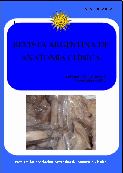
THORAX-ABDOMINAL VAGUS NERVES IN FETUSES. Nervios vagos toraco-abdominales en fetos
DOI:
https://doi.org/10.31051/1852.8023.v7.n3.14188Palabras clave:
Vagus nerves, esophageal innervation, thoracic esophagus, anatomy, surgical anatomy, Nervios vago, inervación del esófago, esófago torácico, anatomía, anatomía quirúrgicaResumen
Los nervios vagos han sido exhaustivamente estudiados en los adultos pero no en los niños, y mayormente en el trayecto intracraneal, más que en la periferia. El objetivo de este estudio fue proveer información más específica sobre los nervios vagos toraco-abdominales, describirlos en fetos y asociarlos con la rotación gástrica, de modo que pueda ser aplicada a procedimientos clínicos, reduciendo la morbilidad. Se disecaron treinta fetos entre 12 y 23 semanas de gestación, mayormente varones (87%), desde la parte inferior del cuello hasta el cardias, identificando los troncos y ramas de los nervios vagos. Los nervios fueron descriptos en su ingreso en el tórax en relación con las arterias carótidas, en su posición en el tercio superior del esófago asociados con el origen de las ramas cardíacas y pulmonares, en el tercio inferior del esófago con muchas variaciones en su distribución, a nivel diafragmático en el hiato esofágico y, finalmente, en relación con la posición gástrica. La discusión involucró descripciones hechas por diferentes autores incluyendo algunos estudios recientes que proporcionan resultados electrofisio-lógicos y consideraciones de aspectos clínicos, principalmente representados por procedimientos quirúrgicos y su morbilidad, ambos asociados con la lesión de los nervios vagos.
Vagus nerves have been extensively studied in adults but not in fetuses, and mostly in the intracranial pathway than the peripheral one. The objective of this study was to provide more specific information on the thorax-abdominal vagus nerves, to describe them in fetuses and to associate them with the gastric rotation, so it could be applied to clinical procedures, reducing morbidity. Thirty fetuses between 12 to 23 weeks of gestation, mainly male (87%), were dissected from the lower neck to the cardias, identifying vagus nerve trunks and braches. Vagus nerves were described at the entrance in the thorax in relation with the carotid arteries, in their position at the upper third of the esophagus associated with the origin of cardiac and pulmonary branches, in the lower third of the esophagus with many variations in their distribution, at the diaphragmatic level in the esophageal hiatus and, finally, in relation with the gastric position. The discussion involved descriptions made by different authors including some recent studies providing electrophysiological results and considerations on clinical aspects, mainly represented by surgical procedures and their morbidity associated, both to vagus nerve injury.
Descargas
Citas
Ezemba N, Eze JC, Nwafor IA, Etukokwu KC, Orakwe OI. 2014. Colon interposition graft for corrosive esophageal stricture: midterm functional outcome. World J Surg 38: 2352-57.
Fragoso AC, Ortiz R, Hernandez F, Olivares P, Martínez L, Tovar JA. 2015. Defective upper gastrointestinal function after repair of combined esophageal and duodenal atresia. J Pediatr Surg 50: 531-34.
Gopal M, Westgarth-Taylor C, Loveland J. 2015. Repair of tracheo-oesophageal fistula secondary to button battery ingestion: A combined cervical and median sternotomy approach. Afr J Paediatr Surg 12: 91-93.
Gratacós E, Gómez R, Nicolaides K, Romero R, Cabero L. 2007. Medicina Fetal. Ed Med Panamericana, Buenos Aires, pp.388.
Kuwahara T, Takahashi A, Takahashi Y, Kobori A, Miyazaki S, Takei A, Fujino T, Okubo K, Takagi K, Fujii A, Takigawa M, Watari Y, Hikita H, Sato A, Aonuma K. 2013. Clinical characteristics and management of peri-esophageal vagal nerve injury complicating left atrial ablation of atrial fibrillation: lessons from eleven cases. J Cardiovasc Electrophysiol 24: 847-51.
Miyake H, Hayashi S, Kawase T, Cho BH, Murakami G, Fujimiya M, Kitano H. 2010. Fetal anatomy of the human carotid sheath and the structures in and around it. Anat Rec (Hoboken) 293: 438-45.
Miyano G, Yamoto M, Morita K, Kaneshiro M, Miyake H, Nouso H, Koyama M, Nakajima H, Fukumoto K, Urushihara N. 2015. Laparoscopic Toupet fundoplication for gastroesophageal reflux: A series of 131 neurologically impaired pediatric cases at a single children’s hospital. Pediatr Surg Int 10: 925-29.
Nebot-Cegarra J, Maraculla-Sanz E, Reina de la Torre F. 1999. Factors involved in the “rotation” of the human embryonic stomach along its longitudinal axis computer-assisted morpho-metrical analysis. J Anat 194: 61-69.
Ortíz R, Galán AS, Martínez L, Domínguez E, Hernández F, Santamaría ML, Tovar AJ. 2015. Tertiary surgery for complicated repair of esophageal atresia. Eur J Pediatr Surg 25: 20-26.
Prives M (Lysenkov N, Bushkovic V). 1985. Anatomía Humana. Tomo II. Min Publishers, Moscu, pp. 307.
Razumosky AY, Alhasov AB, Bataev SH, Yekimosvskaya EV. 2015. Laparoscopic fundoplication Nissen-Gold standard treatment of gastroesophageal reflux in children. Eksp Klin Gastroenterol 1: 72-77.
Sadler TW, Langman J. 2007. Embriología Médica con Orientación Clínica. Ed Med Panamericana, Buenos Aires, pp. 215.
Seki A, Green HR, Lee TD, Hong LS, Tan J, Vinters HV, Chen P-S, Fishbein MC. 2014. Sympathetic nerve fibers in human cervical and thoracic vagus nerves. Heart Rhythm 11: 1411-17.
Spitz L. 2014. Esophageal replacement: over-coming the need. J Pediatr Surg 49: 849-52.
Takassi GF, Herbella FA, Patti MG. 2013. Anatomic variations in the surgical anatomy of the thoracic esophagus and its surrounding structures. Arq Bras Cir Dig 26:101-06.
Tsuboi I, Hayashi M, Miyauchi Y, Iwasaki Y, Yodogawa K, Hayashi H, Uetake S, Takahashi K, Shimizu W. 2014. Anatomical factors associated with periesophageal vagus nerve injury after catheter ablation of atrial fibrillation. Nippon Med Sch 81: 248-57.
Testut L, Latarjet A. 1973. Anatomía Humana. Tomos III – IV. Salvat Ed, Barcelona, pp. (III) 172-77, 983-84, (IV) 253-56.
Verlinden TJ, Rijkers K, Hoogland G, Herrler A. 2015. Morphology of the human cervical vagus nerve: implications for vagus nerve stimulation treatment. Acta Neurol Scand. DOI 10.1111/ane.12462.
Weijs TJ, Ruurda JP, Luyer MD, Nieuwenhuijzen GA, van Hillegersberg R, Bleys RL. 2015. Topography and extent of pulmonary vagus nerve supply with respect to transthoracic oesophagectomy. J Anat 227: 431-39.
Williams PL, Warwick R. 1992. Gray Anatomia. Tomo I. Churchill Livingstone Ed, Madrid, pp. 225, 1185-86.
Yu C, Ramgopal S, Libenson M, Abdelmoumen I, Powell C, Remy K, Madsen JR, Rotenberg A, Loddenkemper T. 2014. Outcome of vagal nerve stimulation in a pediatric population: a single center experience. Seizure 23: 105-11.
Zuidema GD. 1999. Cirugía del Aparato Digestivo. Tomo I. Ed Med Panamericana, Buenos Aires, pp. 17-22.
Descargas
Publicado
Cómo citar
Número
Sección
Licencia
Los autores/as conservarán sus derechos de autor y garantizarán a la revista el derecho de primera publicación de su obra, el cuál estará simultáneamente sujeto a la Licencia de reconocimiento de Creative Commons que permite a terceros compartir la obra siempre que se indique su autor y su primera publicación en esta revista. Su utilización estará restringida a fines no comerciales.
Una vez aceptado el manuscrito para publicación, los autores deberán firmar la cesión de los derechos de impresión a la Asociación Argentina de Anatomía Clínica, a fin de poder editar, publicar y dar la mayor difusión al texto de la contribución.


