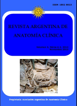
COURSE OF THE MAXILLARY ARTERY THROUGH THE LOOP OF THE AURICULOTEMPORAL NERVE. Recorrido de la arteria maxilar a través del bucle del nervio aurículotemporal
DOI:
https://doi.org/10.31051/1852.8023.v5.n3.14081Palabras clave:
Auriculotemporal nerve, maxillary artery, variations, Nervio aurículotemporal, arteria maxilar, variacionesResumen
Las variaciones en el curso de la arteria maxilar se describen a menudo, con sus relaciones con el músculo pterigoideo lateral. En el presente caso informamos una variación exclusiva en el curso de la arteria maxilar que no fue publicada antes. En un cadáver masculino de 75 años arteria maxilar derecho estaba pasando por el bucle del nervio auriculo-temporal. La arteria meníngea media provenía de la arteria maxilar con un bucle del nervio auriculo-temporal. La arteria maxilar pasaba profunda con respecto al nervio dentario inferior pero superficial al nervio lingual. El conocimiento de estas variaciones es importante para el cirujano y también serviría para explicar la posible participación de estas variaciones en la etiología del dolor mandibular.
Variations in the course of the maxillary artery are often described with its relations to the lateral pterygoid muscle. In the present case we report a unique variation in the course of the maxillary artery which was not reported before. In a 75 years old male cadaver the right maxillary artery passed through the loop of the auriculotemporal nerve. The middle meningeal artery was arising from the maxillary artery within the nerve loop of auriculotemporal nerve. Further the maxillary artery passed deep to the inferior alveolar nerve but superficial to the lingual nerve. The knowledge of these variations is important for surgeons and it would also explain the possible involvement of these variations in etiology of the craniomandibular pain.
Referencias
Bergman RA, Adel K. Afifi, AK, Miyauchi R. 1988. Illustrated Encyclopedia of Human Anatomic Variation: Opus II: Cardiovascular System: Arteries: Head, Neck, and Thorax. Urban and Schwarzenberg. http://www.anatomyatlases.org/AnatomicVariants/Cardiovascular/Text/Arteries/Maxillary.html
Blermann H. 1943. DeChirugische-Bedeetung der Lage variationen de Arteria Maxillaris. Anat Anz 94:289-309
Claire PG, Gibbs K, Hwang SH, Hill RV. 2011. Divided and reunited maxillary artery: developmental and clinical considerations. Anat Sci Int 86: 232-236.
Gulekon N, Anil A, Poyraz A, Peker T, Turgut HB, Karakose M. 2005.Variations in the anatomy of the auriculotemporal nerve. Clnc Anat 18: 15-22.
Hussain A, Binahmed A, Karim A, Sándor GKB. 2008. Relationship of the maxillary artery and lateral pterygoid muscle in a Caucasian sample. Oral Surg Oral Med Oral Pathol Oral Radiol Endod 105: 32-36.
Kolotyluk DR. 1997. Mechanical stenosis of the maxillary artery in the infratemporal fossa: An etiology of craniomandibular pain and vascular headache. Department of Radiology and Diagnostic Imaging Edmonton: University of Albert. www.collectionscanada.gc.ca/obj/.pdf. Last accessed on 12/ 03/ 2013.
Kwak HH, Jo JB, Hu Ks, Oh CS, Koh Ks, Chung IH. 2010. Topography of the third portion of the maxillary artery via the transantral approach in Asians. J craniofacsurg, 21: 1284-1289.
Namking M, Boonruangsri P, Woraputtaporn W, Guldner FH. 1994. Short Report Communic-ation between the facial and auriculotemporal nerves. J Anal, 185: 421-426.
Otake, Ippeikageyama, Ikuomataga, Izumi. 2011. Clinical anatomy of the maxillary artery. Okajimas folia anatomica Japonica 87: 154-164.
Racz L, Maros T, Seres-Sturm L. 1981. Anatomical variations of the nervus alveolaris inferior and their importance for the practice. ANAT ANZ 149: 329-332.
Standring S. 2008. Gray's Anatomy. 40thedn, Spain: Elsevier Limited, 540-1.
Uysal II, Buyukmumcu M, Dogan NU, Seker M, Ziylan T. 2011. Clinical Significance of Maxillary Artery and its Branches: A Cadaver Study and Review of the Literature. International Journal of Morphology 29: 1274.
Descargas
Publicado
Número
Sección
Licencia
Los autores/as conservarán sus derechos de autor y garantizarán a la revista el derecho de primera publicación de su obra, el cuál estará simultáneamente sujeto a la Licencia de reconocimiento de Creative Commons que permite a terceros compartir la obra siempre que se indique su autor y su primera publicación en esta revista. Su utilización estará restringida a fines no comerciales.
Una vez aceptado el manuscrito para publicación, los autores deberán firmar la cesión de los derechos de impresión a la Asociación Argentina de Anatomía Clínica, a fin de poder editar, publicar y dar la mayor difusión al texto de la contribución.


