
Anatomical study of the pancreatic ducts and duodenal papillae.
DOI:
https://doi.org/10.31051/1852.8023.v14.n2.37158Keywords:
Duodenum; Pancreas; Pancreatic duct; Duodenal papilla; Anatomy.Abstract
Abstract:
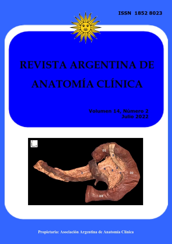
Objectives: The pancreatic ducts and duodenal papillae present variable location and morphology. This variability must be known when performing endoscopic, surgical or radiological procedures that involve them; necessary in the diagnosis and treatment of biliodigestive pathologies. The objective of the present study is to review the anatomy of these structures, record possible variations and carry a review of the literature.Material and method: A descriptive, observational anatomical study was carried out, in which 40 duodenum-pancreatic blocks were dissected from adult corpses of both sex preserved in formaldehyde. We recorded the frequency, location and morphology of both duodenal papillae, the prevalence of the main and accessory pancreatic ducts and the site of junction between the two ducts. These findings were compared with classic and recent literature.
Results: The percentage relative frequency of the major and minor duodenal papillae was 100% and 87.5% respectively. The first was predominantly located at the second portion of the duodenum, in its middle third and in the posteromedial sector (82.5%); while the minor papilla was found in the upper third of the second duodenal portion, antero-medial sector (80%). The main and accessory pancreatic ducts were found in 97.5% and 87.5% of the cases, respectively. The junction of the pancreatic ducts was found in 54.3%.
Conclusions: Classical anatomy prevailed in the studied structures.
Downloads
References
Avisse C, Flament J, Delattre J. 2000. Ampulla of vater Anatomic, Embryologic, and Surgical Aspects. Surg Clin North Am 80: 201-12.
Belou P. 1915. Anatomía de los conductos biliares y de la arteria cítica. Buenos Aires. Oceana. 60, 270.
Classen M, Hellwig H, Rosch W. 1973. Anatomy of the Pancreatic duct. A duodenoscopic-radiological study. Endoscopy 5: 14.
Costamagna G, Nuzzo G, Puglionisi A. 1982. Le variazioni di implanto duodenale della papilla di Vater. Minerva Chir 37: 1573.
Dimitriou I, Katsourakis A, Nikolaidou E, Noussios G. 2018. The Main Anatomical Variations of the Pancreatic Duct System: Review of the Literature and Its Importance in Surgical Practice. J Clin Med Res 10: 370-75.
Dowdy G, Waldron G, Brown W. 1962. Surgical Anatomy of the Pancreatobiliary Ductal System. Arch Surg 84: 229-46.
Estapé G, Boudrandi S, Veirano G, Cardozo R, Carriquiry G, Neirotti R. 1985. Anatomía quirúrgica de la papila duodenal. Montevideo. Oficina del Libro. 59-69.
Flati G, Flati D, Porowska B,Ventura T, Catarci M, Carboni M. 1994. Surgical anatomy of the papilla of Vater and biliopancreatic ducts. Am Surg 60: 712-18.
Ignatov M, Zahariev A, Pose S, Olivera E. 2021. Reporte de dos casos de variantes de los conductos pancreáticos. Rev Argent Anat Online 12: 95-98.
Jirasiritham J, WilasrusmeeC, Poprom N, Larbcharoensub N. 2016.Pancreaticobiliary Ductal Anatomy in the Normal Population.Asian Pac J Cancer Prev 17: 4363-65.
Li L, Yamataka A, Wang Y, Wang D, Wang K, Li Z, Shimizu T, Yamashiro Y, Zhang J, Lane G, Miyano T. 2003. Anomalous pancreatic duct anatomy, ectopic distal location of the papilla of Vater and congenital biliary dilatation: a new developmental triad?. Pediatr Surg Int 19: 180-85.
Poppel MH, Jacobson HG. 1956. Roentgen Aspects of the Papilla of Vater. Am J Dig Dis 1: 49-58.
Rouviere H, Delmas A. 2005.Anatomía Humana: descriptiva, topográfica y funcional. Tomo 2. 11 ª Edición. Barcelona: Masson S.A. 418.
Testut JL, Latarjet A. 1984. Tratado de Anatomía Humana. Tomo 4. 9 ª Edición. Barcelona: Salvat editores. 670.
Wilasrusmee C, Pongchairerks P. 1999. Pancreaticobiliary ductal anatomy in Thai People. J Hepatobiliary Pancreat Surg 1: 79-85.
Downloads
Published
How to Cite
Issue
Section
License
Copyright (c) 2022 María F. Ignatov Galan, Santiago Pose Veirano, Alexander M. Zahariev Marrero

This work is licensed under a Creative Commons Attribution-NonCommercial 4.0 International License.
Authors retain copyright and grant the journal right of first publication with the work simultaneously licensed under a Creative Commons Attribution License that allows others to share the work with an acknowledgement of the work's authorship and initial publication in this journal. Use restricted to non commercial purposes.
Once the manuscript has been accepted for publications, authors will sign a Copyright Transfer Agreement to let the “Asociación Argentina de Anatomía Clínica” (Argentine Association of Clinical Anatomy) to edit, publish and disseminate the contribution.

