
Anatomo-tomographic study of the acetabular versión
DOI:
https://doi.org/10.31051/1852.8023.v12.n3.30218Keywords:
acetabular versión; CT (computer tomography); retroversion.Abstract
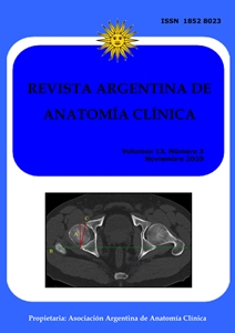
Introduction: The imaging study of the morphometric characteristics of the hip joint, in a sample of asymptomatic cases, is important to understand variations from normality. Materials and method: This is a retrospective study on CT scans requested because of abdominal-pelvic pathologies, in both genders, aged between 18 and 45 years, orthopedi-cally healthy. A digital measurement of the acetabular version was performed. Variables considered and analyzed with descriptive statistics were age, sex and acetabular version. Results: 165 pelvises were included, 102 male (61.9%) and 63 female (38.1%). The mean age of the cohort was 36.4 years. The acetabular version was positive (anteversion) in 163 cases, with a mean male of 17.1° ± 2.7° (p <0.03) and female of 19.3° ± 3.9° (p <0.01). Two male pelvises presented retroversion (1.2%). The difference in the contralateral version mean was 1.15° ± 2.2°. Conclusion: The female acetabulum has a higher average anteversion than the male. Different contralateral acetabular anteversion is common. The prevalence of retroversion in an asymptomatic young adult sample is low.
Downloads
References
Anda S, Svenningsen S, Grontvedt T, Benum P. 1990. Pelvic inclination and spatial orientation of the acetabulum. A radiographic, computed tomographic and clinical investigation. Acta Radiol 31: 389-94.
Ganz R, Parvizi J, Beck M, Leunig M, Notzli H, Siebenrock KA. 2003. Femoroacetabular impingement: a cause for osteoarthritis of the hip. Clin Orthop Relat Res 417: 112-20.
Giori NJ, Trousdale RT. 2003. Acetabular retroversion is associated with osteoarthritis of the hip. Clin Orthop Relat Res 417: 263-69.
Jamali AA, Deuel C, Perreira A, Salgado CJ, Hunter JC, Strong EB. 2007. Linear and angular measurements of computer-generated models: are they accurate, valid, and reliable? Comput Aided Surg 12: 278-85.
Jamali AA, Mladenov K, Meyer DC, Martinez A, Beck M, Ganz R, Leunig M. 2007. Antero-posterior pelvic radiographs to assess acetabular retroversion: high validity of the “cross-over-sign” J Orthop Res 25: 758-65.
Kalberer F, Sierra RJ, Madan SS, Ganz R, Leunig M. 2008. Ischial spine projection into the pelvis: a new sign for acetabular retroversion. Clin Orthop Relat Res 466: 677-83.
Klasan A, Neri T, Sommer C, Leie MA, Dworschak P, Schofer MD, Heyse TJ. 2019. Analysis of acetabular version: Retroversion prevalence, age, side and gender correlations. J Orthop Translat 18: 7-12.
Klingenstein GG, Zbeda RM, Bedi A, Magennis E, Kelly BT. 2013. Prevalence and preoperative demographic and radiographic predictors of bilateral femoroacetabular impingement. Am J Sports Med 41: 762-68.
Larson CM, Moreau-Gaudry A, Kelly BT, Byrd JW, Tonetti J, Lavallee S. 2015. Are normal hips being labeled as pathologic? A CT-based method for defining normal acetabular coverage. Clin Orthop Relat Res 473: 1247-54.
Maruyama M, Feinberg JR, Capello WN, D’Antonio JA. 2001. The Frank Stinchfield Award: Morphologic features of the acetabulum and femur: anteversion angle and implant positioning. Clin Orthop Relat Res 393: 52-65.
Murtha PE, Hafez MA, Jaramaz B, DiGioia AM. 2008. Variations in acetabular anatomy with reference to total hip replacement. J Bone Joint Surg Br 90: 308-13.
Perreira AC, Hunter JC, Laird T, Jamali AA. 2011. Multilevel measurement of acetabular version using 3-D CT-generated models: implications for hip preservation surgery. Clin Orthop Relat Res 469: 552-61.
Rubalcava J, Gómez-García F, Ríos-Reina JL. 2012. Ángulo de anteversión acetabular de la cadera en población adulta mexicana medida por tomografía computada. Acta Ortop Mex 26: 155-61.
Sengodan VC, Sinmayanantham E, Kumar JS. 2017. Anthropometric analysis of the hip joint in South Indian population using computed tomography. Indian J Orthop 51: 155-61.
Siebenrock KA, Schoeniger R, Ganz R. 2003. Anterior femoro-acetabular impingement due to acetabular retroversion: treatment with periacetabular osteotomy. J Bone Joint Surg Am 85: 278-86.
Stem ES, O’Connor MI, Kransdorf MJ, Crook J. 2006. Computed tomography analysis of acetabular anteversion and abduction. Skeletal Radiol 35: 385-89.
Suzuki D., Nagoya S., Takashima H., Tateda K., Yamashita T. 2017. Three-dimensional orienta-tion of the acetabulum. Clin Anat 30: 753-60.
Tallroth K, Lepistö J. 2006. Computed tomo-graphy measurement of acetabular dimensions: Normal values for correction of displasia. Acta Orthop 77: 598-602
Tannenbaum E, Kopydlowski N, Smith M, Bedi A, Sekiya JK. 2014. Gender and racial differences in focal and global acetabular version. J Arthroplasty 29: 373-76.
Vandenbussche E, Saffarini M, Taillieu F, Mutschler C. 2008. The asymmetric profile of the acetabulum. Clin Orthop Relat Res 466: 417-23.
Wassilew GI, Heller MO, Janz V, Perka C, Müller M, Renner L. 2017. High prevalence of acetabular retroversion in asymptomatic adults: a 3D CT-based study. Bone Joint J 99-B: 1584-89.
Werner CM, Copeland CE, Ruckstuhl T, Stromberg J, Turen CH, Kalberer F. 2010. Radiographic markers of acetabular retro-version: correlation of the cross-over sign, ischial spine sign and posterior wall sign. Acta Orthop Belg 76: 166-73.
Zeng Y, Wang Y, Zhu Z, Tang T, Dai K, Qiu S. 2012. Differences in acetabular morphology related to side and sex in a Chinese population. J Anat 220: 256-62.
Zhang H, Wang Y, Ai S, Chen X, Wang L, Dai K. 2017. Three-dimensional acetabular orientation measurement in a reliable coordinate system among one hundred Chinese. PloS one, 12, e0172297.
Downloads
Published
How to Cite
Issue
Section
License
Copyright (c) 2020 Carlos M. Quinteros

This work is licensed under a Creative Commons Attribution-NonCommercial 4.0 International License.
Authors retain copyright and grant the journal right of first publication with the work simultaneously licensed under a Creative Commons Attribution License that allows others to share the work with an acknowledgement of the work's authorship and initial publication in this journal. Use restricted to non commercial purposes.
Once the manuscript has been accepted for publications, authors will sign a Copyright Transfer Agreement to let the “Asociación Argentina de Anatomía Clínica” (Argentine Association of Clinical Anatomy) to edit, publish and disseminate the contribution.

