A NOVEL ANOMALY OF THE ANTERIOR DIGASTRIC MUSCLE. UNA ANOMALÍA NUEVA DEL MÚSCULO DIGÁSTRICO ANTERIOR
DOI:
https://doi.org/10.31051/1852.8023.v2.n3.13893Palabras clave:
neck, radiology, variation, cuello, anatomía, variacíon anatómicaResumen
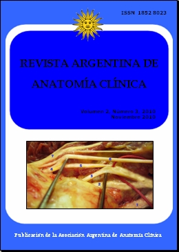
Documentamos una anomalía del músculo digástrico anterior en un cadáver femenino de 85 años de edad. La anomalía consiste en cuatro vientres adicionales que salen entre los dos típicos digástricos anterior. Analizamos el significado embriológico y clínico de esta variante. El músculo digástrico es derivado de dos arcos branquiales o arcos faríngeos. El primer arco branquial forma el vientre anterior y el segundo forma el vientre posterior. Actuando juntos, los vientres ayudan a impulsar hacia abajo la mandíbula y a estabilizar el hioides. Varias anormalidades en el vientre anterior del digástrico han sido previamente descriptas en la literatura, pero ninguna ha replicado la formación precisa descripta aquí.
We document a novel anomaly of the anterior digastrics of an 85 year old female cadaver, consisting of four additional muscle bellies existing between the two typical anterior digastrics and go on to explain the embryologic and clinical significance of the variant. The digastric muscle is derived from two pharyngeal arches, the first pharyngeal arch forming the anterior belly and the second forming the posterior belly. Acting together, both bellies help to depress the mandible and stabilize the hyoid. Several abnormalities in the anterior belly of the digastric muscle have previously been described in the literature, but none have replicated the precise formation described here.
Referencias
Celik H, Yilmaz E, Atasever A, Durgun B, & Taner D. 1992. Bilateral anatomical anomaly of anterior bellies of digastric muscles. Kaibogaku Zasshi.Journal of Anatomy, 67: 650-651.
Heude E, Bouhali K, Kurihara Y, Kurihara H, Couly G, Janvier P, Levi G. 2010. Jaw muscularization requires Dlx expression by cranial neural crest cells. Proceedings of the National Academy of Sciences, 107: 11441-11446.
Larsson S G, Lufkin R B. 1987. Anomalies of digastric muscles: CT and MR demonstration. Journal of Computer Assisted Tomography, 11: 422-425.
Loukas M, Louis R G, Kapos T, Kwiatkowska M. 2005. A case of a bilateral accessory digastric muscle. Folia Morphologica, 64: 233-236.
Macalister A. 1882: cited in Michna H. 1989. Anatomical anomaly of human digastric muscle. Acta Anatomica 134: 263-264.
Mancuso A A, Harnsberger H R, Muraki A S, Stevens M H. 1983. Computed tomography of cervical and retropharyngeal lymph nodes: Normal anatomy, variants of normal and applications in staging head and neck cancer. part II: Pathology. Radiology, 148: 715-723.
Mashkevich G, Wang J, Rawnsley J, Keller G S. 2009. The utility of ultrasound in the evaluation of submental fullness in aging necks. Archives of Facial Plastic Surgery, 11: 240-245.
Moore K L, Persaud T V N. 2003. Before We Are Born: Essentials of Embryology and Birth Defects (sixth edition). Saunders Elsevier Science USA, Philadelphia, PA. Chapter 11.
Muraki A. S., Mancuso A. A., Harnsberger H. R., Johnson, L. P., Meads G. B. 1983. CT of the oropharynx, tongue base, and floor of the mouth: Normal anatomy and range of variations, and applications in staging carcinoma. Radiology, 148: 725-731.
Peker T., Turgut H. B., Anil A. 2000. Bilateral anomaly of anterior bellies of digastric muscles. Surgical and Radiologic Anatomy, 22: 119-121.
Sadler T.W. 1995. Langman's Medical Embryology (seventh edition). Williams Wilkins, Baltimore, MD. Chapter 16.
Sargon M. F., Onderoglu S., Surucu H. S., Bayramoglu A., Demiryurek D. D., Ozturk H. 1999. Anatomic study of complex anomalies of the digastric muscle and review of the literature. Okajimas Folia Anatomica Japonica, 75: 305-313.
Som P. M. 1987. Lymph nodes of the neck. Radiology, 165: 593-600.
Tank PW. 2005. Grant’s Dissector. Thirteenth Edition. Baltimore: Lippincott Williams & Wilkins, 1-219.
Traini M. 1983. Bilateral accessory digastric muscles. Anatomia Clinica, 5: 199-200.
Descargas
Publicado
Número
Sección
Licencia
Los autores/as conservarán sus derechos de autor y garantizarán a la revista el derecho de primera publicación de su obra, el cuál estará simultáneamente sujeto a la Licencia de reconocimiento de Creative Commons que permite a terceros compartir la obra siempre que se indique su autor y su primera publicación en esta revista. Su utilización estará restringida a fines no comerciales.
Una vez aceptado el manuscrito para publicación, los autores deberán firmar la cesión de los derechos de impresión a la Asociación Argentina de Anatomía Clínica, a fin de poder editar, publicar y dar la mayor difusión al texto de la contribución.


