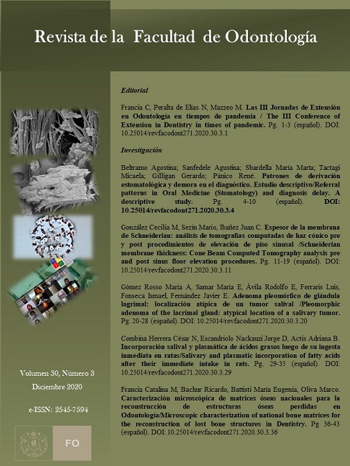
Caracterización microscópica de matrices óseas nacionales para la reconstrucción de estructuras óseas perdidas en Odontología
Palabras clave:
microscopia electrónica de barrido, matriz ósea, rehabilitaciónResumen
Objetivo. caracterizar morfológicamente la ultra estructura de algunas matrices óseas de fabricación nacional disponibles en el mercado comercial local a fin de determinar las principales particularidades que permitan su uso en la rehabilitación odontológica de los rebordes óseos atróficos. Métodos. Se estudiaron matrices óseas provenientes de Hueso Humano Liofilizado (HHL) provistas por el Laboratorio de Hemoderivados de la Universidad Nacional de Córdoba, con formato de bloque y de polvo cuyo tamaño de partícula es de 200 a 1000 μm y en forma de gránulos finos de 1000 a 2000 μm. Las muestras se metalizaron respectivamente con una capa de oro o cromo y fueron analizadas en un microscopio electrónico de barrido (MEB). Resultados. Se observó una estructura del hueso HHL similar a los observados en los espacios del sistema de vasos sanguíneos Haversiano del hueso humano normal. Conclusiones. Los hallazgos estructurales del hueso HHL, similares a los observados en los espacios del sistema de vasos sanguíneos Haversiano del hueso humano normal, garantizan la conservación topográfica responsable del incremento del área superficial y consecuentemente de una gran relación superficie-volumen que favorece la reparación y regeneración ósea constituyendo características favorables para ser utilizadas como injertos. Además, el mantenimiento de la nanoestructura de la matriz extracelular le otorga mayor rugosidad, permitiendo un mayor anclaje que favorece un mejor crecimiento celular por presentación de señales topográficas esenciales, beneficiando la adhesión, proliferación y diferenciación de las células.
Referencias
1. Gómez de Ferraris ME, Campos Muñoz A. Periodoncio de Inserción: Cemento, Ligamento Periodontal y Hueso Alveolar. En: Histología y Embriología Bucodental. Ed Médica Panamericana. Madrid, 2002: 2ª ed., pp 368-83.
2. Hof M, Tepper G, Semo B, Arnhart C, Watzek G, Pommer B. Patients’ perspectives on dental implant and bone graft surgery: questionnaire-based interview survey. Clin. Oral Impl. Res., 2012, 1–4.
3. Misch Carl E. Rationale for Dental Implants- Anatomical Consequences of Edentulism. En: Dental Implant Prosthetics. Elsevier Health Sciences, 2014: 2 ª ed., pp 10-23.Traini et al, 2015
4. Brekke J, Toth J. Principles of Tissue Engineering Applied to Programmable Osteogenesis. J. Biomed. Mater. Res. (Appl Biomater), 1998, 43:380-98.
5. Weibrich G, Götz H, Gnoth S-H, Trettin R, Duschner H, Wagner W. Charakterisierung der Oberflächenmorphologie von Knochenersatzmaterialienmittels REM. Z Zahnärztl Implantol: 2000: 16: 151-159.
6. ThaKral GK, ThaKral Rashmi, Sharma N, Jyotsana S, Vashish P. Nano surface – The Future of Implants. Journal of Clinical and Diagnostic Research. 2014 May, Vol-8(5): 7-10]
7. Rani, V.V.; Vinoth-Kumar, L.; Anitha, V.C.; Manzoor, K.; Deepthy, M.; Shantikumar, V.N. Osteointegration of titanium implant is sensitive to specific nanostructure morphology. Acta Biomater. 2012, 8, 1976–1989.
8. Wang, X.; Gittens, R.A.; Song, R.; Tannenbaum, R.; Olivares-Navarrete, R.; Schwartz, Z.; Chen, H.; Boyan, B.D. Effects of structural properties of electrospun TiO2 nanofiber meshes on their osteogenic potential. Acta Biomater. 2012, 8, 878–885.
9. Perán M, García M A, Lopez-Ruiz E, Jiménez G, and Marchal JA. How Can Nanotechnology Help to Repair the Body? Advances in Cardiac, Skin, Bone, Cartilage, and Nerve Tissue Regeneration. Materials 2013, 6(4), 1333-1359.
10. Alpaslan E, Webster T J. Nanotechnology and pico technology to increase tissue growth: a summary of in vivo studies. Short report. International Journal of Nanomedicine 2014:9 (Suppl 1) 7–12.
11. Kearney J.N. Sterilization on human tissue implants, British Asociation of Tissue Bank 1996 Newsletter 6, 2-5.
12. Bailey A.J. Effect of ionizing radiation on connective tissue components. En: International Review of Connective Tissue Research, D.A. 1968. Hall, ed. Academic Press. New York vol 4.
13. Houben J.C. Free radical produced by ionizing radiation in bone and its constituents. International Journal of Radiation Biology. 1971, 20: 373-389.
14. Dziedzig-Goclawska A. Aplicación de la radiación ionizante para esterilizar aloinjertos de tejido conectivo. En: radiación y operación de Banco de Tejidos, ed. Printing Services AC. Peru, 2002.
Descargas
Publicado
Número
Sección
Licencia

Esta obra está bajo una licencia internacional Creative Commons Atribución-NoComercial-CompartirIgual 4.0.
Aquellos autores/as que tengan publicaciones con esta revista, aceptan los términos siguientes:
- Los autores/as conservarán sus derechos de autor y garantizarán a la revista el derecho de primera publicación de su obra, el cuál estará simultáneamente sujeto a la Licencia de reconocimiento de Creative Commons que permite a terceros:
- Compartir — copiar y redistribuir el material en cualquier medio o formato
- La licenciante no puede revocar estas libertades en tanto usted siga los términos de la licencia
- Los autores/as podrán adoptar otros acuerdos de licencia no exclusiva de distribución de la versión de la obra publicada (p. ej.: depositarla en un archivo telemático institucional o publicarla en un volumen monográfico) siempre que se indique la publicación inicial en esta revista.
- Se permite y recomienda a los autores/as difundir su obra a través de Internet (p. ej.: en archivos telemáticos institucionales o en su página web) después del su publicación en la revista, lo cual puede producir intercambios interesantes y aumentar las citas de la obra publicada. (Véase El efecto del acceso abierto).

