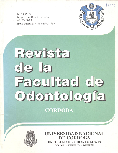
Aspectos estructurales de los dientes primarios. Estudio al M.O. Y M.E.B.
Keywords:
Histology, Tooth, Deciduous, Microscopy, Scanning ProbeAbstract
Most of the histological studies made on deciduous teeth haveonly referred to the estructural aspects of sorne of their tissues, in relation to the exfolation and to the general or specific dental pathology. Therefore, we aimed at an integral study of hard tissues in primary molars to be used in future investigations of clinical application. For this reason it was our purpose, in this first stage, to make an integral study of the hard tissues of the primary molars. Thirty clinically molars investigated, twenty- six showed a normal clinic and four molars presented the "white lesion". Polishing technique was used for obsertations on the optic microscopy while sectioning was perfonned for scanning electron microscopy (SEM). The following findings of this research were: a) Enarnel, prismless externa! zone and neonatal line and microdefects were easily identified. Few an little noticeable Retzius stries can be observed in the posnatallayer. However adamantine spindles and penetrating tubules are abundan!.
b) Chance in the direction of tubules proximal lo the pulp charnber !loor and absence of peritubular dentine is outstanding.
e) In30% ~f the analyzed molars fissures or hypominemlized area> were evindenced al intraradicular leve!. The Czermak spaces are not frequent. d) Acelular cementum prevails. e) The molars with the white lesion exhibit the histological characteristic of the dental cariousDownloads
Published
Issue
Section
License
Aquellos autores/as que tengan publicaciones con esta revista, aceptan los términos siguientes:
- Los autores/as conservarán sus derechos de autor y garantizarán a la revista el derecho de primera publicación de su obra, el cuál estará simultáneamente sujeto a la Licencia de reconocimiento de Creative Commons que permite a terceros:
- Compartir — copiar y redistribuir el material en cualquier medio o formato
- La licenciante no puede revocar estas libertades en tanto usted siga los términos de la licencia
- Los autores/as podrán adoptar otros acuerdos de licencia no exclusiva de distribución de la versión de la obra publicada (p. ej.: depositarla en un archivo telemático institucional o publicarla en un volumen monográfico) siempre que se indique la publicación inicial en esta revista.
- Se permite y recomienda a los autores/as difundir su obra a través de Internet (p. ej.: en archivos telemáticos institucionales o en su página web) después del su publicación en la revista, lo cual puede producir intercambios interesantes y aumentar las citas de la obra publicada. (Véase El efecto del acceso abierto).

