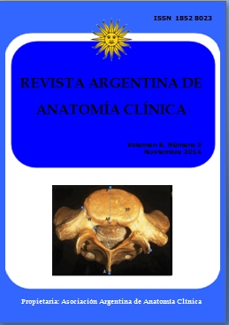
MORPHOMETRY OF THE PEDICLE OF FIRST SACRAL VERTEBRAE AND ITS APPLICATION IN POSTERIOR TRANSPEDICULAR SCREW FIXATION. Morfometría del pedículo de la primera vértebra sacra y su aplicación en la fijación posterior con tornillo transpedicular
DOI:
https://doi.org/10.31051/1852.8023.v6.n3.14140Palabras clave:
pedicle, sacrum, spine, screw, vertebra, Pedículo, sacro, columna vertebral, tornillo, vértebraResumen
Los objetivos del presente estudio fueron determinar los parámetros anatómicos del pedículo S1 en la población India del sur para comparar los datos con respecto a los géneros masculinos y femeninos. El estudio incluyó 50 sacros secos (25 hombres y 25 mujeres) que se obtuvieron en el laboratorio de anatomía de nuestra institución. En el presente estudio se observa que la longitud media del pedículo S1 fue 49.9± 3,6 mm para los hombres y 46.3± 4,8 mm para las mujeres. La altura céfalo-caudal del pedículo S1 fue 27.2±4.0 mm y 23.9±3.7 mm para el varón y la hembra respectivamente. La anchura antero-posterior del pedículo S1 fue 7.5± 1,3 mm, 7.5± 1.7 mm en varones y mujeres, respectivamente. La distancia antero-posterior de S1, desde el promontorio sacro a la apófisis espinosa de S1 fue 52.9± 5.2 mm y 50.4± 6.8 mm en los géneros masculino y femenino respectivamente. El presente estudio demostró que la longitud y la altura de céfalo-caudal eran más altos (p0.05) en varones que en mujeres. Los datos de mujeres y varones con respecto a la anchura antero-posterior y la distancia antero-posterior de S1 no eran estadísticamente diferentes. El presente estudio ha proporcionado datos morfométricos importantes del pedículo de la primera vértebra sacra de la muestra anatómica de la población India del sur. El conocimiento de los diámetros del pedículo de S1 es crucial para la colocación segura de tornillos para la fijación transpedicular posterior.
Objectives of the present study were to determine the anatomical parameters of the S1 pedicle in South Indian population and to compare the data, with respect to male and female genders. The study included 50 dry sacra (25 male and 25 female), which were obtained from the anatomy laboratory of our institution. It is observed in the present study that the mean S1 pedicle length was 49.9± 3.6 mm for male and 46.3± 4.8 mm for the female. The cephalocaudal heights of S1 pedicle were 27.2±4.0 mms and 23.9±3.7 mms for the male and female respectively. The anteroposterior width of S1 pedicle was 7.5± 1.3 mms, 7.5± 1.7 mms in males and females respectively. The anteroposterior distances of S1, from the sacral promontory to the spinous process of S1 were 52.9± 5.2 mms and 50.4± 6.8 mms respectively for the male and female genders. The present study observed that the mean S1 pedicle length and the cephalocaudal height were higher (p<0.05) for the males than that of females. The data (male vs female) were not found statistically different (p>0.05), with respect to the anteroposterior width of the S1 pedicle and the anteroposterior distances of S1 from the sacral promontory to the spinous process of S1. The present study has provided important morphometric data onto the pedicle of the first sacral vertebrae, from the anatomical samples of the South Indian population. The knowledge of pedicle diameters of S1 is crucial to the safe placement of screws in the posterior transpedicular screw fixation.
Referencias
Arman C, Naderi S, Kiray A, Aksu FT, Y?lmaz HS, Tetik S, Korman E. 2009. The human sacrum and safe approaches for screw placement. J Clin Neurosc 16: 1046-49.
Bogduk N. 2005. Clinical anatomy of the lumbar spine and sacrum. 4th edn. London: Churchill Livingstone, 59.
Brantley AG, Mayfield JK, Koeneman JB, Clark KR. 1994. The effects of pedicle screw fit. An in vitro study. Spine 19: 1752-58.
Diel J, Ortiz O, Losada RA, Price DB, Hayt MW, Katz DS. 2001. The sacrum: pathologic spectrum, multimodality imaging, and sub-specialty approach. Radiographics 21: 83-104.
Ebraheim NA, Xu R, Biyani A, Nadaud MC. 1997. Morphologic considerations of the first sacral pedicle for iliosacral screw placement. Spine 22: 841-46.
Esses SI, Botsford DJ, Huler RJ, Rauschning W. 1991. Surgical anatomy of the sacrum: A guide for rational screw fixation. Spine 16: S283–88.
Frymoyer JW. 1988. Back pain and sciatica. N Engl J Med 318: 291–300.
Harrington PR, Dickson JH. 1976. Spinal instrum-entation in the treatment of severe progressive spondylolisthesis. Clin Orthop 117: 157-63.
Kostuik JP. 1986. Techniques of internal fixation for degenerative conditions of the spine. Clin Orthop 203: 219-31.
Krogman WM, Iscan MY. 1986. The Human Skeleton in Forensic Medicine. 2nd edn. Springfield, Illinois: Charles C Thomas.
Lonstein JE, Denis F, Perra JH, Pinto MR, Smith MD, Winter RB. 1999. Complications assoc-iated with pedicle screws. J Bone Joint Surg Am 81: 1519-28.
Misenhimer GR, Peek RD, Wiltse LL, Rothman SLG, Widell EH. 1989. Anatomic analysis of pedicle cortical and cancellous diameter as related to screw size. Spine 11: 367-72.
Morales-Ávalos R, Leyva-Villegas JI, Vílchez-Cavazos F, Martínez-Ponce de León ÁR, Elizondo-Omaña RE, Guzmán-López S. 2012. Morphometric characteristics of the sacrum in Mexican population. Its importance in lumbo-sacral fusion and fixation procedures. Cir Cir 80: 528-35.
Morales-Avalos R, Re Elizondo-Omaña RE, Vílchez-Cavazos F, Martínez-Ponce de León AR, Elizondo-Riojas G, Delgado-Brito M, Cortés -González P, Guzmán-Avilán RI, Pinales-Razo R, de la Garza-Castro O, Guzmán-López S. 2012. Vertebral fixation with a transpedicular approach. Relevance of anatomical and imaging studies. Acta Ortop Mex 26: 402-11.
Okutan O, Kaptanoglu E, Solaroglu I, Beskonakli E, Tekdemir I. 2003. Pedicle morphology of the first sacral vertebra. Neuroanatomy 2: 16-19.
Okutan O, Kaptanoglu E, Solaroglu I, Beskonakli E, Tekdemir I. 2004. Determination of the length of anteromedial screw trajectory by measuring interforaminal distance in the first sacral vertebra. Spine 29: 1608-11.
Peretz AM, Hipp JA, Heggeness MH. 1998. The internal bony architecture of the sacrum. Spine 23: 971-974.
Vij K. 2011. Textbook of Forensic Medicine and Toxicology. Principles and Practice. 5th edn. New Delhi: Elsevier.
Xu R, Ebraheim NA, Yeasting RA, Wong FY, Jackson WT. 1995. Morphometric evaluation of the first sacral vertebra and the projection of its pedicle on the posterior aspect of the sacrum. Spine 20: 936-40.
Descargas
Publicado
Número
Sección
Licencia
Los autores/as conservarán sus derechos de autor y garantizarán a la revista el derecho de primera publicación de su obra, el cuál estará simultáneamente sujeto a la Licencia de reconocimiento de Creative Commons que permite a terceros compartir la obra siempre que se indique su autor y su primera publicación en esta revista. Su utilización estará restringida a fines no comerciales.
Una vez aceptado el manuscrito para publicación, los autores deberán firmar la cesión de los derechos de impresión a la Asociación Argentina de Anatomía Clínica, a fin de poder editar, publicar y dar la mayor difusión al texto de la contribución.


