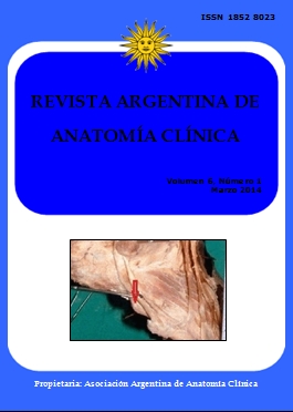
COMPARATIVE ANATOMICAL STUDY AND INCIDENCE OF OS PERONEUM IN PERONEUS LONGUS TENDON AND ITS CLINICAL SIGNIFICANCE. Estudio anatómico comparativo e incidencia del os peroneum en el tendón de peroneo largo y su significación clínica
DOI:
https://doi.org/10.31051/1852.8023.v6.n1.14093Palabras clave:
Os peroneum, sesamoid bone, Jones fracture, styloid, Huesos sesamoideos, Fractura de Jones, EstiloidesResumen
Introducción: El objetivo de nuestro estudio fue evaluar la tasa de incidencia del os peroneo (OP) en el tendón del peroneo lateral largo (PLT) y su importancia clínica. Métodos: La disección de 60 cadáveres embalsamados (56 hombres y 4 mujeres) del grupo de mediana edad se hizo para tener acceso a la tasa de incidencia del os peroneo en PLT. Resultados: En nuestro estudio se observó que la tasa de incidencia del os peroneo fue de 86,6% (52 extremidades). La ubicación del os peroneo es también un tema de controversia. La mayoría de los autores afirman que se relaciona con el hueso cuboides y de vez en cuando se ve inferior al calcáneo distal a la articulación calcáneo-cuboidea. Pero en el presente estudio el os- peroneo estuvo en relación al hueso cuboides en 40 extremidades (76.9%) y distal a articulación calcaneocuboidea en el resto de las 12 extremidades (23.1%). Conclusión: Este estudio sugiere que existe una alta tasa de incidencia de un OP en cadaveres. Esto puede ser como consecuencia de la técnica utilizada para localizar el mismo. La importancia clínica ha sido mencionada en relación con la ubicación del os peroneo, que puede ser confundido con fracturas estiloides y de Jones.
Introduction: The aim of our study was to assess the incidence rate of the os peroneum (OP) in the peroneus longus tendon (PLT) and its clinical significance. Methods: Dissection of 60 embalmed cadavers (56 male and 4 female) of middle age group was done to access the incidence rate of os peroneum in peroneus longus tendon. Results: In our study the incidence rate of os peroneum was 86.6% (52 limbs). The location of os peroneum is also a subject of dispute. Most authors stated that it is related to the cuboid bone and occasionally it is seen inferior to the calcaneum distal to the calcaneocuboid joint. But in the present study os peroneum was in relation to cuboid bone in 40 limbs (76.9%) and distal to calcaneocuboid joint in 12 limbs (23.1%). Conclusion: This study suggests that there is a high incidence rate of the os peroneum in the peroneus longus tendon in cadavers. This may be a consequence of the technique used to locate it. The os peroneum can be mistaken for a styloid or Jones fractures.
Referencias
Benninger B, Kloenne J. 2011. The Clinical Importance of the Os Peroneum: A Dissection of 156 Limbs Comparing the Incidence Rates in Cadavers versus Chronological Roentgen-ograms. The Foot and Ankle Online Journal 4: 2.
Bhargava KN, Sanyal P Bhargava SN. 1961. Lateral musculature of the leg as seen in hundred Indian Cadavers. Indian Journal of Medical Science 15: 181-185.
Bizarro AH.. 1921. On sesamoid and super-numerary bones of the limbs. J Anat 55: 256–268.
Bloom RA. 1991. The infracalcaneal os peroneum. Acta Anat 140: 34-36.
Carter DR, Orr TE, Fyhrie DP, Schurman DJ. 1987. Influences of mechanical stress on prenatal and postnatal skeletal development. Clin Orthop 219: 237–250.
Carter DR, Wong M, Orr TE. 1991. Musculoskeletal ontogeny, phylogeny, and functional adaptation. J Biomech 24: 3–16.
Carter DR. 1987. Mechanical loading history and skeletal biology. J Biomech 20: 1095–1109.
Goldberg I, Nathan H. 1987. Anatomy and pathology of the sesamoid bones. Int Orthop 11: 141–147.
Jones FW. 1942. Principles of anatomy as seen in the hand. Baltimore: Williams and Wilkins. p 203–207.
LeMinor JM. 1987. Comparative anatomy and significance of the sesamoid bone of the peroneus longus muscle (os peroneum). J Anat 151: 85–99.
Maurer M, Lehrman J. 2012. Significance of Sesamoid Ossification in Peroneus Longus Tendon Ruptures. J Foot Ankle Surg 51: 352-355.
Mellado JM, Ramos A, Salvadó E, Camins A, Danús M, Saurí A. 2003. Accessory ossicles and sesamoid bones of the ankle and foot: imaging findings, clinical significance and differential diagnosis. Eur Radiol 13: L164-L177.
Merida-Velasco JA, Sanchez-Montesinos I, Espin-Ferra J, Rodriguez-Vazquez JF, Merida-Velasco JR, Jiminez-Collado J. 1997. Development of the human knee joint. Anat Rec 248: 269–278.
Muehleman C, Williams J, Bareither ML. 2009. A radiologic and histologic study of the os peroneum: prevalence, morphology, and relationship to degenerative joint disease of the foot and ankle in a cadaveric sample. Clin Anat 22: 747-754.
Ogden JA. 1984. Radiology of postnatal skeletal development. X. Patella and tibial tuberosity. Skeletal Radiol 11: 246–257.
Oydele O, Maseko C, Mkasi N, Mashanyana M. 2006 High incidence of the os peroneum in a cadaver sample in Johannesburg, South Africa: possible clinical implication?. Clin Anat 19: 605-610.
Pancoast HK. 1909. Radiographic statistics of the sesamoid in the tendon of the gastrocnemius. U Penn Med Bull 22: 213–217.
Pearson K, Davin AG. 1921. On the sesamoids of the knee joint. I. Man. II. Evolution of the sesamoids. Biometrika 13: 133–175, 350–400.
Rühli FJ, Solomon LB, Henneberg M. 2003. High prevalence of tarsal coalitions and tarsal joint variants in recent cadavers sample and its possible significance. Clin Anat 16: 411-415.
Sarin VK, Erickson GM, Giori NJ, Bergman AG, Carter DR. 1999. Coincident development of sesamoid bones and clues to their evolution. Anat Rec 15; 257: 174-80.
Small KM, Potter SS. 1993. Homeotic transformations and limb defects in Hox A11 mutant mice. Genes Dev 7: 2318–2328.
Standring S. 2005. Greys Anatomy. The Anatomical Basis of clinical practice.40th Ed.Philadelphia; p 1420.
Storm EE, Kingsley DM. 1996. Joint patterning defects caused by single and double mutations in members of the BMP family. Development 122: 3969–3979.
Stropeni L 1920. Frattura isolate di un osso soprannumerario del tarso (os peroneum externum).. Arch.ital.d.chir 2: 56.
Descargas
Publicado
Número
Sección
Licencia
Los autores/as conservarán sus derechos de autor y garantizarán a la revista el derecho de primera publicación de su obra, el cuál estará simultáneamente sujeto a la Licencia de reconocimiento de Creative Commons que permite a terceros compartir la obra siempre que se indique su autor y su primera publicación en esta revista. Su utilización estará restringida a fines no comerciales.
Una vez aceptado el manuscrito para publicación, los autores deberán firmar la cesión de los derechos de impresión a la Asociación Argentina de Anatomía Clínica, a fin de poder editar, publicar y dar la mayor difusión al texto de la contribución.


