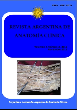
POSSIBLE ENTRAPMENT OF THE ULNAR ARTERY BY THE THIRD HEAD OF PRONATOR TERES MUSCLE. El posible atrapamiento de la arteria ulnar por el tercer fascículo del músculo pronador teres
DOI:
https://doi.org/10.31051/1852.8023.v4.n3.14035Palabras clave:
Ulnar artery, brachial artery, third head of pronator teres, entrapment, arteria cubital, arteria braquial, tercer fascículo del pronador teres, atrapamientoResumen
El conocimiento de las variaciones en los alrededores de la fosa cubital es útil para cirujanos ortopédicos, cirujanos plásticos y médicos en general. Observamos las variaciones arteriales y musculares en y alrededor de la fosa cubital. La arteria braquial terminó 2 pulgadas por encima de la base de la fosa cubital. Las arterias radiales y cubitales entraron en la fosa cubital pasando delante de los tendones de los músculos braquial y bíceps braquial respectivamente. La arteria cubital estaba rodeada por el tercer fascículo del pronador teres, que tuvo su origen en la fascia cubriendo la parte distal del músculo braquial. Este músculo se unió a tendón de pronador teres distalmente y fue suministrado por una rama del nervio mediano. Este músculo podría alterar el flujo sanguíneo en la arteria cubital y puede causar dificultades para el registro de la presión sanguínea.
Knowledge of variations at and in the surroundings of cubital fossa is useful for the orthopedic surgeons, plastic surgeons and medical practitioners in general. During routine dissection, we observed arterial and muscular variations in and around the cubital fossa. The brachial artery terminated 2 inches above the base of the cubital fossa. The radial and ulnar arteries entered the cubital fossa by passing in front of the tendons of brachialis and biceps brachii respectively. The ulnar artery was surrounded by the third head of pronator teres which took its origin from the fascia covering the distal part of the brachialis muscle. This muscle joined pronator teres tendon distally and was supplied by a branch of median nerve. This muscle could alter the blood flow in the ulnar artery and may cause difficulties in recording the blood pressure.
Referencias
Biswas S, Adhigari A, Kundu P. 2010. Variations in the cubital fossa. International Journal of Anatomical Variations (IJAV) 3: 122.
Celik HH, Gormus G, Aldur MM, Ozcelik M. 2001. Origin of the radial and ulnar arteries: variations in 81 arteriograms. Morphologie 85: 25-27.
Cho KO. 1978. Entrapment occlusion of the ulnar artery in the hand. A case report. J Bone Joint Surg Am 60: 841-843.
Ciervo A, Kahn M, Pangilinan AJ, Dardik H. 2001. Absence of the brachial artery: report of a rare human variation and review of upper extremity arterial anomalies. J Vasc Surg 33: 191-194.
Dartnell J, Sekaran P, Ellis H. 2007. The superficial ulnar artery: incidence and calibre in 95 cadaveric specimens. Clin Anat 20: 929-932.
Datta AK. 1997. The arm. In: Essentials of human anatomy, part III. 2nd Ed Current books international. Calcutta, 62-63.
George BM, Nayak SB. 2008. Median nerve and brachial artery entrapment in the abnormal brachialis muscle-A case report. Neuroanatomy 7: 41–42.
Jelev L, Georgiev GP. 2009. Unusual high-origin of the pronator teres muscle from a Struthers' ligament coexisting with a variation of the musculocutaneous nerve. Rom J Morphol Embryol. 50: 497-499.
Le Double AF. 1897. Traite des variations musculaire de l’homme et de leur signification an point de l’anthropologie zoologique, vol. 1. Paris: Librarie C, Reinwald, Schleicher Freres, 79-82.
Macalister A. 1875. Additional observations on muscular anomalies in human anatomy (3rd series) with a catalogue of the principal muscular variations hitherto published. Transactions of the Royal Irish Academy, 25: 1-134.
Mc Cormack LJ, Caldwell EW, Anson BJ. 1953. Brachial and antebrachial patterns. Surg Gynecol Obst 96: 43-54.
Nayak SR, Krishnamurthy A, Prabhu LV, Jiji PJ, Ramanathan L, Kumar S. 2008. Multiple supernumerary muscles of the arm and its clinical significance. Bratisl Lek Listy 109: 74-75.
Okoro IO, Jiburum BC. 2003. Rare high origin of the radial artery: A bilateral symmetrical case. The Nigerian journal of surgical research 5: 70-72.
Salmons S. 1995. Muscle. In: Williams PL (ed). Gray’s Anatomy. 38th Ed. Edinburgh: Churchill Livingstone, 846.
Senanayake KJ, Salgado S, Rathnayake MJ, Fernando R, Somarathne K. 2007. A rare variant of the superficial ulnar artery, and its clinical implications: a case report. J Med Case Reports 1: 128.
Thane GD. 1892. Arthrology-Myology-Angiology. In: Schafer EA, Thane GD (eds). Quain’s Elements of Anatomy. 10th Ed. London: Longmans, Green and Co, 223-224, 439-441.
Vollala VR, Nagabhooshana S, Bhat SM. 2008. Trifurcation of brachial artery with variant course of radial artery: Rare observation. Anatomical Science International 83: 307–309.
Yoshinaga K, Tanii I, Kodama K. 2003. Superficial brachial artery crossing over the ulnar and median nerves from posterior to anterior: embryological significance. Anat Sci Int. 78: 177-180.
Descargas
Publicado
Número
Sección
Licencia
Los autores/as conservarán sus derechos de autor y garantizarán a la revista el derecho de primera publicación de su obra, el cuál estará simultáneamente sujeto a la Licencia de reconocimiento de Creative Commons que permite a terceros compartir la obra siempre que se indique su autor y su primera publicación en esta revista. Su utilización estará restringida a fines no comerciales.
Una vez aceptado el manuscrito para publicación, los autores deberán firmar la cesión de los derechos de impresión a la Asociación Argentina de Anatomía Clínica, a fin de poder editar, publicar y dar la mayor difusión al texto de la contribución.


